





A Minute of Shaking, Then an Unexpected Diagnosis
A rice farmer in the Philippines was going about her day without any sign of illness – no headache, no confusion, no weakness – when her right arm and leg began shaking uncontrollably. The episode lasted roughly one minute. Then it stopped, and she went to the emergency department.
Doctors ran a physical exam and found nothing unusual. The answers came only after they scanned her brain.
What the Scan Revealed
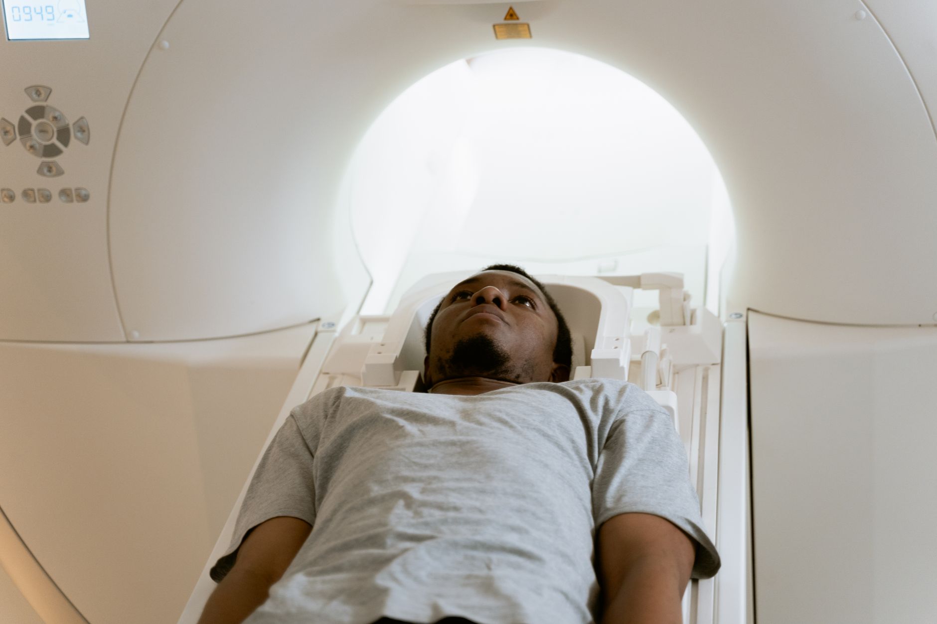
Magnetic resonance imaging of the woman’s left frontal lobe showed a bright cluster of nodules surrounded by fluid. The imaging was distinctive enough that doctors formed their suspicion immediately: those nodules looked like worm eggs embedded in brain tissue. The case was later published in the New England Journal of Medicine, where the attending physicians detailed their diagnostic process and the chain of reasoning that led them from a seizure in a farm worker to a parasitic infection of the central nervous system.
Their working diagnosis was neuroschistosomiasis – a rare and serious complication arising from infection with a Schistosoma parasitic worm, more commonly known as a blood fluke. The condition develops when fluke eggs travel through the bloodstream and lodge in the brain rather than passing through the body’s usual filtration systems. It is considered an uncommon outcome even among people who carry the parasite, which is part of what makes this case medically notable enough for a major journal.
The specific species suspected in her case was S. japonicum, the blood fluke most frequently found in the Philippines. Despite carrying Japan’s name, S. japonicum has actually been eliminated from Japan itself and currently circulates in parts of China and sections of Indonesia, in addition to the Philippines. The naming reflects an older geography of the parasite’s range that no longer holds.
How Lab Work Closed the Case
Diagnosis in neuroschistosomiasis cases doesn’t rest on imaging alone. The confirmatory step – the one that locked in the identification – came from laboratory testing in which the eggs grew tails, a biological characteristic that distinguishes Schistosoma species at a cellular level. That morphological detail, eggs extending into a tailed form under lab conditions, gave physicians the hard confirmation that MRI could only suggest.

The case sits in a category of medical events that illustrates how much diagnostic technology matters when symptoms are brief and physical examination yields nothing. A seizure lasting one minute, in a patient who otherwise appeared well, could point toward dozens of different causes. Without MRI, the nodular cluster in her frontal lobe would have gone undetected. Without the lab confirmation, the specific parasite responsible might have remained uncertain. The entire diagnostic chain depended on layering one tool on top of another – imaging followed by biological testing – until the picture became unambiguous.
The Biology Behind the Infection
Blood flukes are flatworms that live inside the blood vessels of their hosts. S. japonicum typically enters the human body through skin contact with freshwater where infected snails are present – a common occupational exposure for rice farmers, who work in standing water regularly. Once inside the host, the worms reproduce and release eggs. Most eggs exit the body through waste, but a fraction travel the wrong direction, carried by blood flow into tissues where they cause inflammation and, over time, structural damage.
When eggs reach the brain, the immune response around them produces the kind of swelling and nodule formation visible on the woman’s MRI. The left frontal lobe cluster that appeared in her scan represented that inflammatory response at work – her body reacting to foreign material in tissue that is not equipped to expel it. That process, playing out silently until it crossed some threshold, produced the 60-second seizure that sent her to the emergency department in the first place.
Schistosomiasis as a general infection affects hundreds of millions of people globally, but the neurological form remains a distinct and less-studied subset. Most people infected with blood flukes never experience brain involvement at all. The pathway from water exposure to a bright cluster of nodules visible on an MRI in a Manila emergency department involves a specific and relatively unlikely sequence of biological events that researchers are still working to fully characterize.

What the New England Journal of Medicine case report adds to that picture is a documented example of the full diagnostic arc – from an otherwise healthy patient with a brief focal seizure, through imaging, through laboratory confirmation, to a species-level identification. The woman was a rice farmer. The worm was S. japonicum. The eggs grew tails. And none of that was visible from the outside when she walked into the emergency room.